Patent Ductus Arteriosus in a Puppy
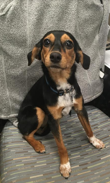
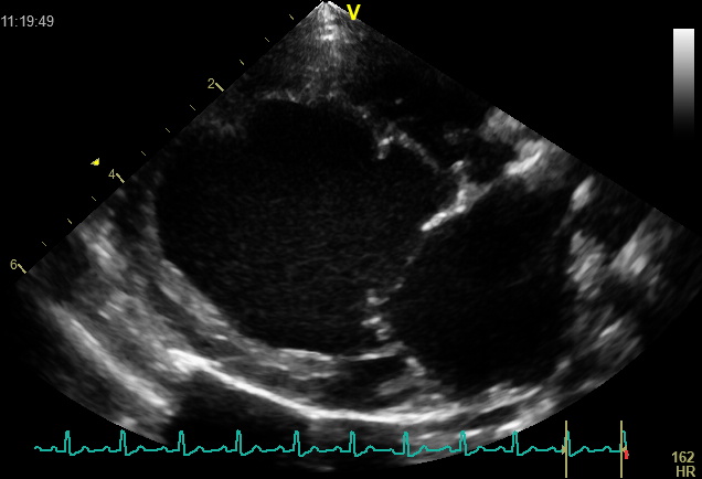
Lady presented to Friendship Cardiology Specialists for evaluation of a heart murmur noted by her vet at her first puppy exam. An echocardiogram (ultrasound of the heart) was performed, and she was diagnosed with a patent ductus arteriosus (PDA) causing severe heart enlargement.
A PDA is a birth defect involving the main blood vessels arising from the heart. The ductus arteriosus is a fetal communication between the aorta and pulmonary artery which should normally close at birth by constriction of muscle around the ductus. Some dogs are born without this muscle, and therefore their ductus cannot close at birth. Failure of closure is called a PDA. The PDA causes extra blood to circulate through the lungs, and overloads the heart’s pumping ability.
Most puppies with a PDA show no symptoms, but a PDA produces a characteristic murmur that can be detected when listening to the chest with a stethoscope. An ultrasound of the heart (echocardiogram) performed by a cardiologist is required for diagnosis. If left untreated, the PDA overloads the heart’s pumping ability. This can cause fluid to back-up into the lungs and cause trouble breathing. This fluid is called congestive heart failure (CHF). If a PDA is not corrected, most patients progress to CHF within the first year of life, and prognosis without treatment is poor. Fortunately, there are excellent treatment options to close the ductus. Once the ductus is closed, the overloaded heart can shrink down to normal size. Most dogs do not require long-term medications or follow-up and can live normal life spans.
Treatment options include minimally invasive surgery or traditional surgery. Minimally invasive surgery is performed by a veterinary cardiologist using catheters passed through blood vessels to introduce occlusion devices that block off blood flow through the PDA from the inside. Traditional surgery is performed by a veterinary surgeon who directly visualizes the ductus from the outside of the heart and closes it with sutures. Both methods have similar success rates, and the choice of treatment depends on the size of the patient and the size and shape of the ductus. In Lady’s case, give her small body size and relatively large ductus size, the decision was made to proceed with traditional surgery with the Friendship Surgical Specialists.
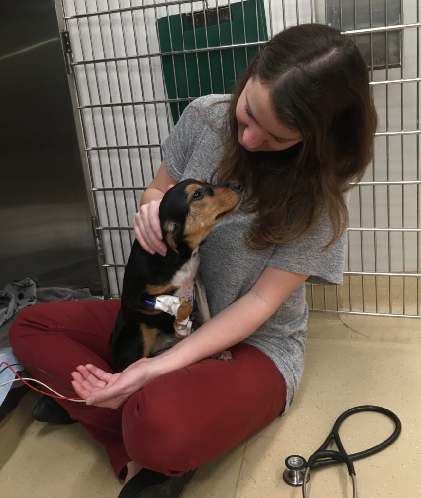
Lady was scheduled for surgery and her owner was instructed to watch for signs of CHF. Soon after her initial appointment, Lady began coughing and having trouble breathing which was concerning for development of CHF. She was started on medications to help prevent fluid build-up and improve the heart’s pumping function and her symptoms improved.
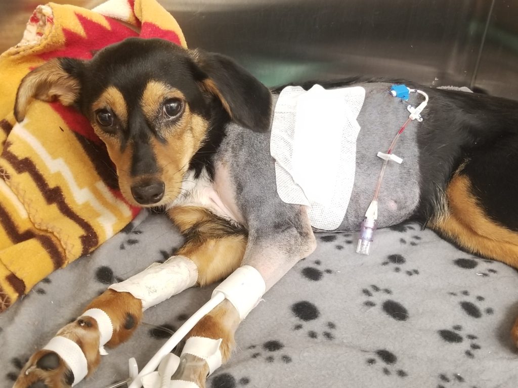
Lady was placed under general anesthesia and the PDA was successfully ligated by specialty surgeon Dr. Mathieu Glassman. A chest tube was placed so that any residual air or fluid could be suctioned from the chest. She recovered well in the intensive care unit where she was treated with pain medications and antibiotics. The following day her chest tube was removed, and she was comfortable and stable for discharge home to her owners.
Lady has an excellent long-term prognosis. She will be seen for a cardiology recheck in two weeks where an echocardiogram will be repeated to ensure the ductus remains closed and her heart size is improving. Her surgeon will assess her incision to make sure she is healing appropriately and remove her stitches. If she is doing well, she will be taken off her heart medications.
Dr. Block earned her veterinary degree from the University of California, Davis, School of Veterinary Medicine in 2013. Dr. Block completed a rotating internship followed by a cardiology residency at the University of Pennsylvania and joined Friendship Cardiology Specialists in 2017.


